Bright Field Microscope Sciencequery

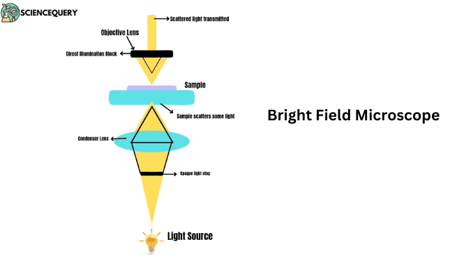
Bright Field Light Microscope Pdf Microscope Laboratory Techniques Bright field microscopy is the simplest of a range of techniques used for illumination of samples in light microscopes, and its simplicity makes it a popular technique. the typical appearance of a bright field microscopy image is a dark sample on a bright background, hence the name. Bright field microscopy is defined as a simple compound microscope technique that uses a condenser lens to focus light on a sample, allowing for the visualization of objects such as living cells and forensic evidence, although it has limitations in contrast, magnification, and resolution.

Bright Field Microscope Sciencequery Brightfield microscope is an optical microscope that uses light rays to produce a dark image against a bright background. it is the standard microscope that is used in biology, cellular biology, and microbiological laboratory studies. brightfield microscope is also known as the compound light microscope. Bright field microscopy – a short introduction right field (or brightfield) illumination with a compound microscope is commonly used when observing specimens. if the contrast of a specimen is sufficient, bright field illumination offers a combination of ease of use and performance. In light microscopy, visible light is used to detect such small objects – with bright field microscopy being the most common form of light microscopy. in bright field microscopy, the image you see is formed mainly by the absorption of light by the specimen (for example a cell) (lackie, 2010). Significance: surgical microscopes provide adjustable magnification, bright illumination, and clear visualization of the surgical field and have been increasingly used in operating rooms.

Bright Field Microscope Sciencequery In light microscopy, visible light is used to detect such small objects – with bright field microscopy being the most common form of light microscopy. in bright field microscopy, the image you see is formed mainly by the absorption of light by the specimen (for example a cell) (lackie, 2010). Significance: surgical microscopes provide adjustable magnification, bright illumination, and clear visualization of the surgical field and have been increasingly used in operating rooms. Bright field light microscopy is an optical imaging technique that uses visible light to create magnified images of small specimens. it illuminates a sample with transmitted white light, producing an image where the specimen appears dark against a bright background. The quantitative light microscopy shared resource at simmons cancer center enables high end optical microscopy to be applied to cancer focused research. Brightfield microscopy, also known as a compound light microscopy, uses light to illuminate a sample and create an image. the sample is placed on a glass slide and illuminated by a light source, typically a halogen lamp or a light emitting diode (led) light.
Comments are closed.