Crowpanel Esp32 Display 1 28 R Inch 240 240 Round Ips Display Capacitive Touch Spi Screen

Crowpanel Esp32 C3 1 28 Inch Round Ips Display Capacitive Touch Spi Sc Robocraze Fying an antibody (e.g., changes in clone, fluorochrome etc.) or adding a new antibody to a panel, the process begins with a selection of an antibody to be included in clinical testing, which involves several considerations. define the purpose of the new antibody in a panel before initiating the design and validation process. antibodies used in. Designed for researchers working with cell suspensions, the protocol ensures optimal detection of surface and internal markers using fluorescently labeled antibodies. it also includes tips for minimizing cell damage and maximizing signal specificity.
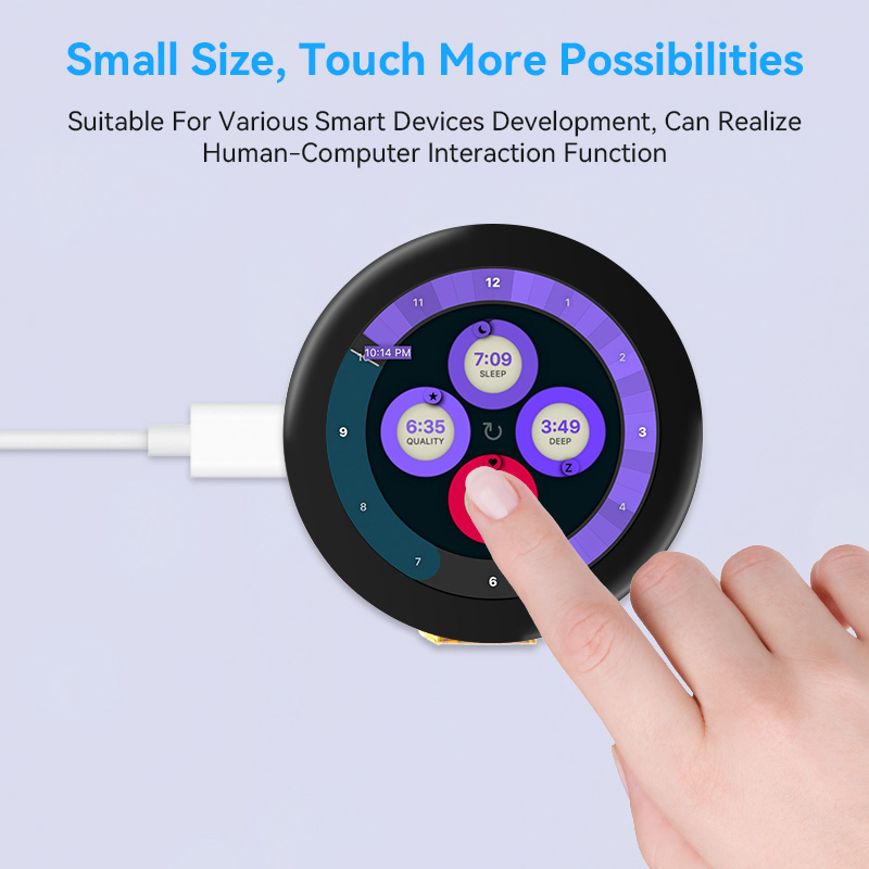
Crowpanel Esp32 Display 1 28 R Inch 240 240 Round Ips Display Capacitive Touch Spi Screen To get the most out of your antibodies, titrate them. Protocols for the staining of intracellular antigens for flow cytometry utilize various fixation and permeabilization methods to allow antibodies access to internal cellular proteins. Flow cytometry typically uses antibodies conjugated to fluorophores to target extracellular markers in order to define cell populations. below, we provide several examples of flow cytometry protocols to choose from depending on cell type, application and target antigen. Combining antibodies designed for live cell phenotyping with antibodies that detect intracellular proteins in flow cytometry can be challenging. cell signaling technology® (cst®) is here to help. this decision tree will enable you to determine the best approach for labeling your targets of interest.
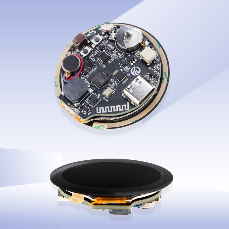
Crowpanel Esp32 Display 1 28 R Inch 240 240 Round Ips Display Capacitive Touch Spi Screen Flow cytometry typically uses antibodies conjugated to fluorophores to target extracellular markers in order to define cell populations. below, we provide several examples of flow cytometry protocols to choose from depending on cell type, application and target antigen. Combining antibodies designed for live cell phenotyping with antibodies that detect intracellular proteins in flow cytometry can be challenging. cell signaling technology® (cst®) is here to help. this decision tree will enable you to determine the best approach for labeling your targets of interest. View our antibody protocol collection to learn more about protein detection in applications including western blot, flow cytometry, and various others. This protocol outlines the key steps involved in performing flow cytometry, highlighting the use of antibodies for cell surface or intracellular marker detection. Dilute primary antibody in fresh blocking permeabilization buffer at the concentration recommended by the antibody supplier. note: you may need to perform a titration experiment to determine the optimal concentration of primary antibody. Flow cytometry protocols for live cells: indirect and direct methods detailed steps to take you through cell preparation and on to indirect and direct methods of flow cytometry. flow cytometry allows you to detect molecules on the cell surface by targeting them with specific antibodies.
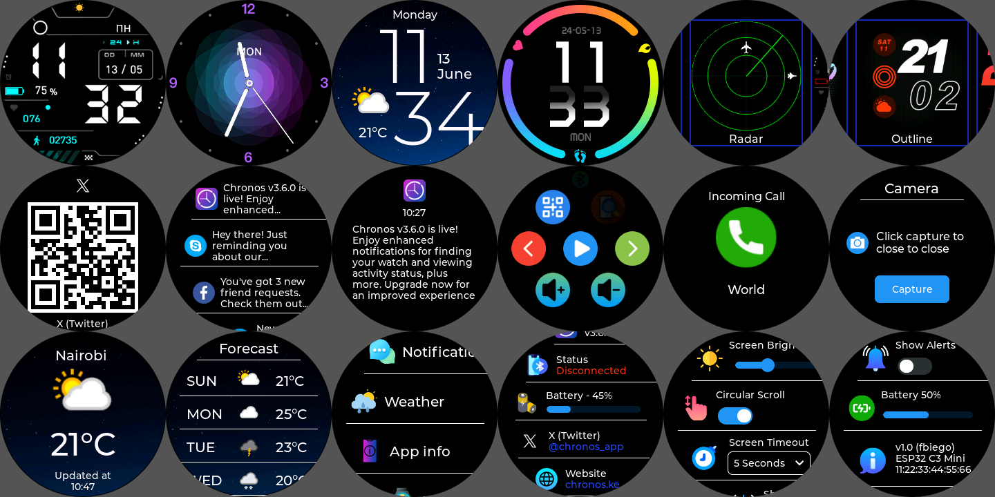
Crowpanel Esp32 Display 1 28 R Inch 240 240 Round Ips Display Capacitive Touch Spi Screen View our antibody protocol collection to learn more about protein detection in applications including western blot, flow cytometry, and various others. This protocol outlines the key steps involved in performing flow cytometry, highlighting the use of antibodies for cell surface or intracellular marker detection. Dilute primary antibody in fresh blocking permeabilization buffer at the concentration recommended by the antibody supplier. note: you may need to perform a titration experiment to determine the optimal concentration of primary antibody. Flow cytometry protocols for live cells: indirect and direct methods detailed steps to take you through cell preparation and on to indirect and direct methods of flow cytometry. flow cytometry allows you to detect molecules on the cell surface by targeting them with specific antibodies.
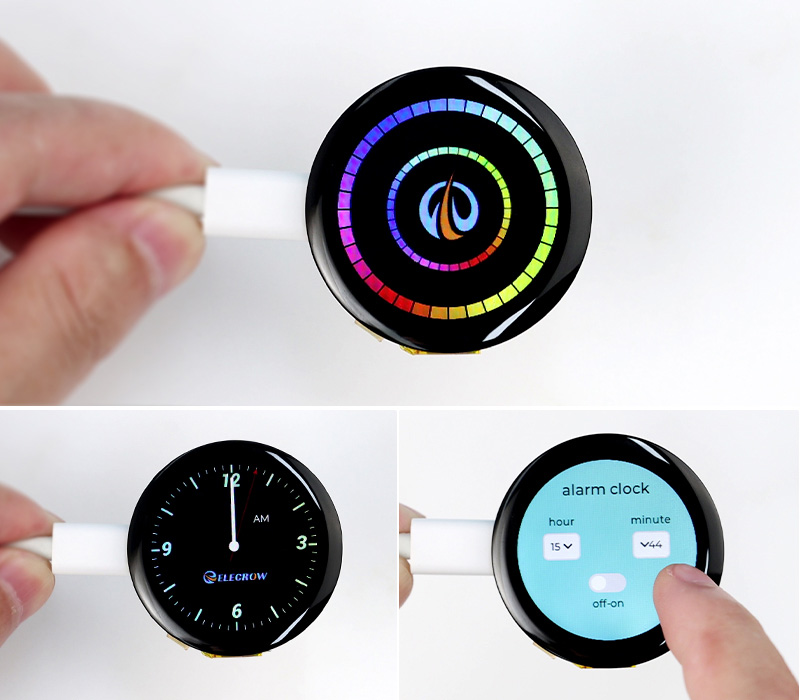
Crowpanel Esp32 Display 1 28 R Inch 240 240 Round Ips Display Capacitive Touch Spi Screen Dilute primary antibody in fresh blocking permeabilization buffer at the concentration recommended by the antibody supplier. note: you may need to perform a titration experiment to determine the optimal concentration of primary antibody. Flow cytometry protocols for live cells: indirect and direct methods detailed steps to take you through cell preparation and on to indirect and direct methods of flow cytometry. flow cytometry allows you to detect molecules on the cell surface by targeting them with specific antibodies.
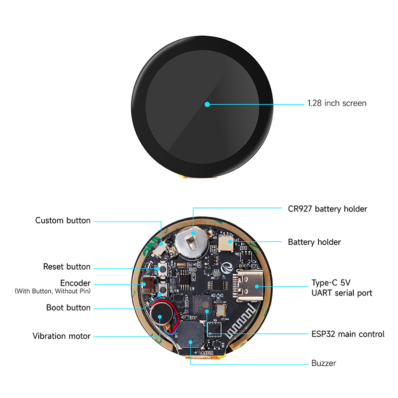
Crowpanel Esp32 Display 1 28 R Inch 240 240 Round Ips Display Capacitive Touch Spi Screen
Comments are closed.