Dermoid Cyst Ultrasound Youtube

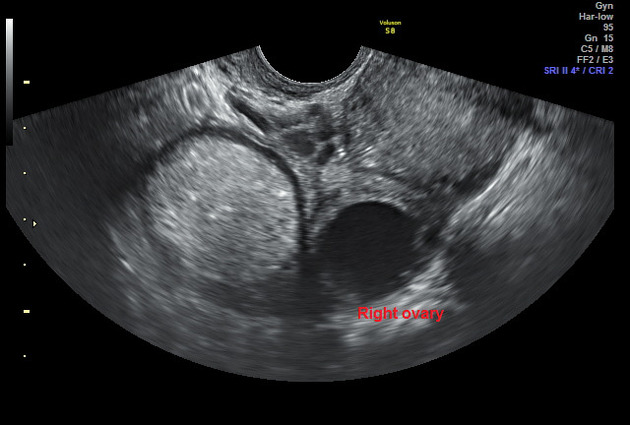
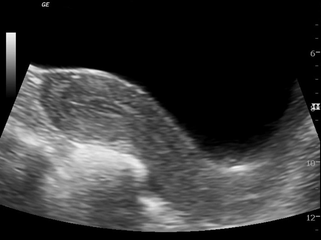
Dermoid Cyst Ultrasound Case 164 57 Off Ultrasound has become a frequently used and highly effective modality through which the diagnosis of a dermoid cyst can be made. a dermoid cyst generally contains fluid, fat and solid tissue. it is this make up that gives rise to the stereotypical sonographic features. On ultrasound, dermoid cysts typically present as well defined, round or oval masses within the ovary. one of their hallmark features is the presence of a mixture of different tissue types, which can create a distinctive appearance.

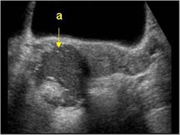
Dermoid Cyst Ultrasound Wikidoc This ultrasound case shows many of the typical features of an ovarian torsion. the ovary is distended with absent doppler flow and some adjacent free fluid. the ovary also contains a 4 cm dermoid cyst with an echogenic rokitansky nodule ('dermoid. In this radiology lecture, we discuss the ultrasound appearance of ovarian dermoid cyst, including the rarely seen but highly specific “floating sphere” sign. Ultrasound cases. home; cases; select category abdomen and retroperitoneum. 1.1 liver 1.2 gallbladder and bile ducts 1.3 pancreas 1.4 spleen 1.5 appendix 1.6 gastrointestinal tract 1.7 peritoneum mesentery and omentum 1.8 various intra abdominal tumors 1.9 retroperitoneum and great vessels 1.10 adrenal glands 1.11 abdominal wall 1.12. Annotated ultrasound images showing the rokitansky nodule, echogenic mesh (hair), dense shadowing foci (tip of the iceberg) and focus of possible tooth ossification which are the characteristic features of an ovarian dermoid cyst. a mature cystic teratoma of the ovary is also called a dermoid cyst.

Dermoid Ovarian Cyst Ultrasound Ultrasound cases. home; cases; select category abdomen and retroperitoneum. 1.1 liver 1.2 gallbladder and bile ducts 1.3 pancreas 1.4 spleen 1.5 appendix 1.6 gastrointestinal tract 1.7 peritoneum mesentery and omentum 1.8 various intra abdominal tumors 1.9 retroperitoneum and great vessels 1.10 adrenal glands 1.11 abdominal wall 1.12. Annotated ultrasound images showing the rokitansky nodule, echogenic mesh (hair), dense shadowing foci (tip of the iceberg) and focus of possible tooth ossification which are the characteristic features of an ovarian dermoid cyst. a mature cystic teratoma of the ovary is also called a dermoid cyst. On ultrasound, dermoid cysts typically present as well defined, cystic masses with heterogeneous internal echoes. a characteristic feature is the presence of echogenic foci representing sebaceous material or hair, often accompanied by posterior acoustic shadowing. On ultrasound, ovarian dermoid cysts appear as cystic lesions with echogenic debris due to the presence of fat, hair, and other tissues. they may also exhibit posterior acoustic shadowing. these benign germ cell tumors can present with pelvic pain or pressure, but they can also be asymptomatic. Dr tahir a siddiqui ( consultant sonologist )ultrasound learning video seriesgujranwala. pakistan0553257350.

Dermoid Ovarian Cyst Ultrasound On ultrasound, dermoid cysts typically present as well defined, cystic masses with heterogeneous internal echoes. a characteristic feature is the presence of echogenic foci representing sebaceous material or hair, often accompanied by posterior acoustic shadowing. On ultrasound, ovarian dermoid cysts appear as cystic lesions with echogenic debris due to the presence of fat, hair, and other tissues. they may also exhibit posterior acoustic shadowing. these benign germ cell tumors can present with pelvic pain or pressure, but they can also be asymptomatic. Dr tahir a siddiqui ( consultant sonologist )ultrasound learning video seriesgujranwala. pakistan0553257350.

Dermoid Cyst Youtube Dr tahir a siddiqui ( consultant sonologist )ultrasound learning video seriesgujranwala. pakistan0553257350.
Comments are closed.