Dna Under Microscope %f0%9f%94%ac%f0%9f%a7%acmicroscope Science Microscopy

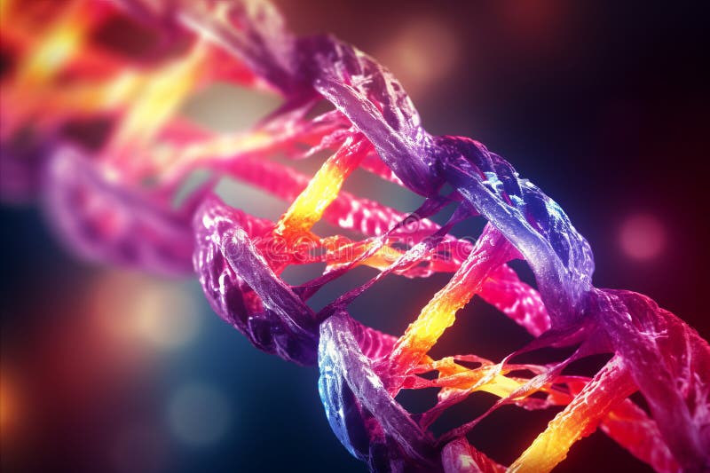
Dna Under Microscope Icon Flat Style Stock Vector Illustration Of Analysis Graphic 130415955 The imaging relies on diffracted light, so when we see an image of the dna’s signature double helix, we’re not really looking at the dna—we see the x rays deflected from atoms. the actual image of dna under a microscope may not be as colorful as our dna illustrations, but it’s realistic. What does dna look like under a microscope? in this informative video, we’ll take you on a journey into the microscopic world of dna. understanding how scien.

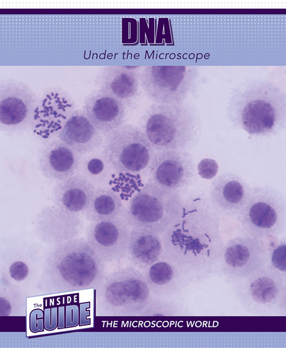
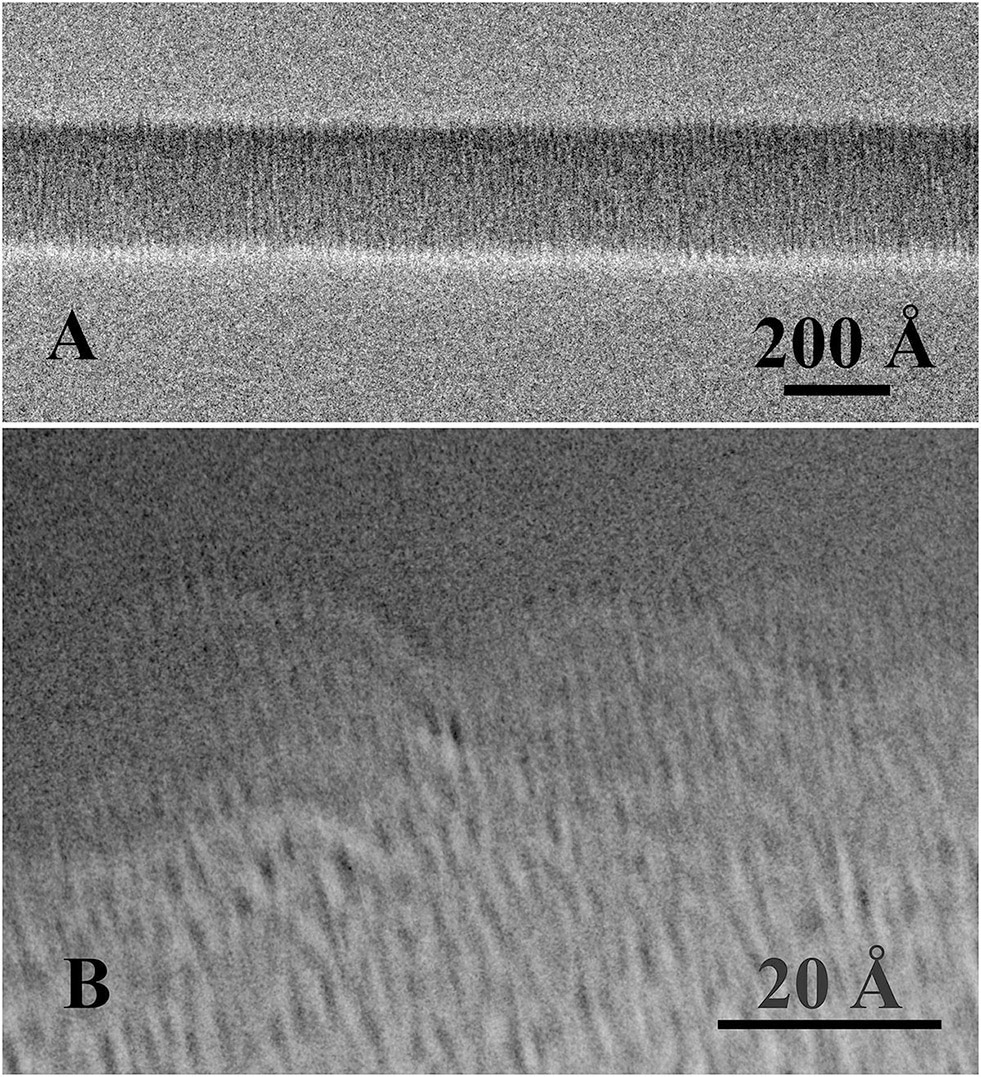
Dna Under The Microscope Cavendish Square Publishing While it is possible to see the nucleus (containing dna) using a light microscope, dna strands threads can only be viewed using microscopes that allow for higher resolution. to view the dna as well as a variety of other protein molecules, an electron microscope is used. We show that through a preparation procedure compatible with the dna physiological conditions, a direct image of a single suspended dna molecule can be obtained. in the image, all relevant lengths of a form dna are measurable. Even though the overall length of a dna molecule is about 2 inches, it is not possible to see dna through light microscopy as the dna is present inside the nucleus inside the cell. dna that has been extracted might be seen through naked eyes as a long thread like structure. Dna cannot be seen in the regular compound microscope. but dna can be seen with the help of more advanced methods like electron microscopy and atomic force microscopy. you can see the nucleus with a compound microscope, but not the dna. chromosomes can be seen only during cell divisions.

Dna Molecule Under The Microscope Generated By Ai Stock Photo Image Of Helix Cytosine 305542308 Even though the overall length of a dna molecule is about 2 inches, it is not possible to see dna through light microscopy as the dna is present inside the nucleus inside the cell. dna that has been extracted might be seen through naked eyes as a long thread like structure. Dna cannot be seen in the regular compound microscope. but dna can be seen with the help of more advanced methods like electron microscopy and atomic force microscopy. you can see the nucleus with a compound microscope, but not the dna. chromosomes can be seen only during cell divisions. Dive into the fascinating world of dna under the microscope. this visual guide explores the techniques, tools, perfect for science lovers. A team of italian scientists have taken a snapshot of a single dna molecule for the first time using the electron microscope. this technological breakthrough comes after years of depending on x ray diffraction to provide indirect, albeit useful, information on the structure of dna. For his ph.d. work, griffith developed the em technology needed to directly visualize bare dna and dna protein complexes. his methods involved carefully controlled rotary shadow casting with tungsten and mounting the dna on very thin carbon films. Under an electron microscope, dna does not appear as the clean, computer generated double helix model. it most often looks like long, thin, and flexible threads. the exact appearance depends on the preparation methods and the type of electron microscope used.

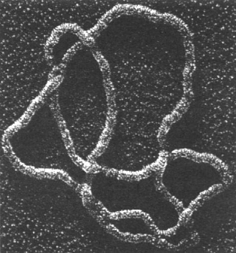
Dna Under Electron Microscope Dive into the fascinating world of dna under the microscope. this visual guide explores the techniques, tools, perfect for science lovers. A team of italian scientists have taken a snapshot of a single dna molecule for the first time using the electron microscope. this technological breakthrough comes after years of depending on x ray diffraction to provide indirect, albeit useful, information on the structure of dna. For his ph.d. work, griffith developed the em technology needed to directly visualize bare dna and dna protein complexes. his methods involved carefully controlled rotary shadow casting with tungsten and mounting the dna on very thin carbon films. Under an electron microscope, dna does not appear as the clean, computer generated double helix model. it most often looks like long, thin, and flexible threads. the exact appearance depends on the preparation methods and the type of electron microscope used.

Dna Under Electron Microscope For his ph.d. work, griffith developed the em technology needed to directly visualize bare dna and dna protein complexes. his methods involved carefully controlled rotary shadow casting with tungsten and mounting the dna on very thin carbon films. Under an electron microscope, dna does not appear as the clean, computer generated double helix model. it most often looks like long, thin, and flexible threads. the exact appearance depends on the preparation methods and the type of electron microscope used.
Comments are closed.