Left Bundle Branch Block Lbbb Ecg Criteria Causes Management The

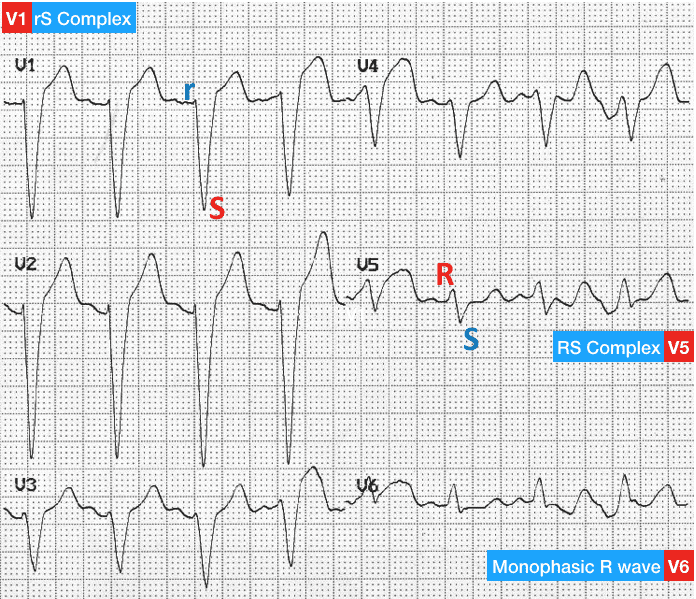
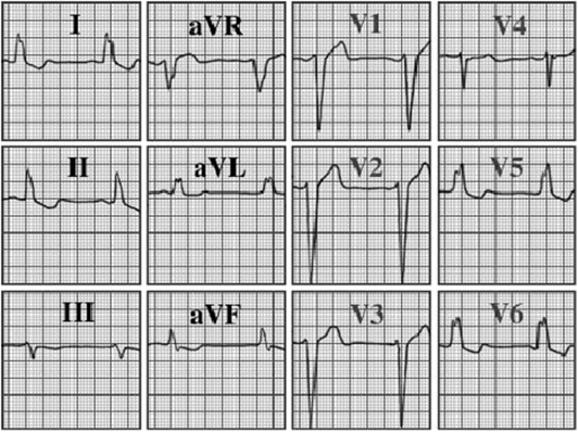
Left Bundle Branch Block Lbbb Ecg Criteria Causes 56 Off Diagnosing left bundle branch block is relatively straightforward. the hallmark of lbbb is the prolonged qrs duration. a qrs duration of 120 ms (0.12 s) or more is required to diagnose a complete left bundle branch block. In patients with significant lv dysfunction, lbbb results in left ventricular dyssynchrony and may contribute to heart failure (hf). the anatomy, clinical manifestations, differential diagnosis, prognostic implications, and treatment of lbbb will be reviewed here.

Left Bundle Branch Block Lbbb Ecg Criteria Causes 51 Off New lbbb in the context of chest pain was once considered a “stemi equivalent” and part of the criteria for thrombolysis. however, more up to date data suggests that chest pain patients with new lbbb have little increased risk of acute myocardial infarction at the time of presentation. Left bundle branch block (lbbb) occurs when something blocks or disrupts the electrical impulse that causes your heart to beat. this block leads to an abnormal heart rhythm. a diagnosis of left bundle branch block often means that you have an underlying heart condition. Identify the clinical features indicative of left bundle branch block. create a clinically guided diagnostic strategy for left bundle branch block. implement individualized management plans for left bundle branch block cases. Left bundle branch block may be due to conduction system degeneration or a reflection of myocardial pathology. left bundle branch block may also develop following aortic valve disease or cardiac procedures.

Left Bundle Branch Block Lbbb Ecg Criteria Causes 51 Off Identify the clinical features indicative of left bundle branch block. create a clinically guided diagnostic strategy for left bundle branch block. implement individualized management plans for left bundle branch block cases. Left bundle branch block may be due to conduction system degeneration or a reflection of myocardial pathology. left bundle branch block may also develop following aortic valve disease or cardiac procedures. Lbbb is supported by mid qrs notching slurring in at least two contiguous leads (especially v5, v6, i, and avl). ( 21376930 ) the most specific finding for lbbb is t ime to qrs notch in lead i is >75 ms (figure below). As the problem is below the atria, the p waves and the pr intervals are normal. the diagnostic criteria for rbbb are: 2. the william marrow mnemonic can be used to quickly recognise left and right bundle branch blocks by looking at v1 and v6:. Right bundle branch block (rbbb) and left bundle branch block (lbbb) both result in delayed ventricular depolarization but differ in their ecg presentations. rbbb typically shows an 'rsr’ pattern in lead v1, while lbbb shows a wide, slurred r wave in leads i, v5, and v6. Explore lbbb causes, heart vector impact, and ecg findings. learn about lbbb in leads v1 v6, septal q waves, ventricular septal infarction, and variants.
Comments are closed.