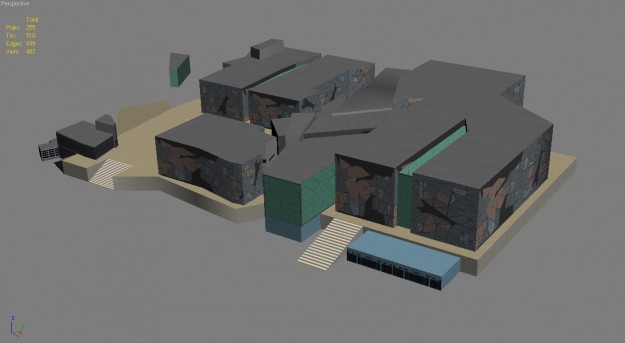
Melbourne Federation Square 3d Model From Cgtrader Com

Melbourne Federation Square 3d Model Cgtrader The appearance of brightness in an ultrasound image is related to how sound waves reflect off different tissues. different tissue densities and compositions cause varying degrees of reflection – or ‘echoing.’ hyperechoic areas strongly reflect sound waves, creating that bright appearance. it’s crucial to remember that “hyperechoic” isn’t a diagnosis; it’s a description of what. The condition is characterized by the presence of bright areas on an ultrasound image of the kidneys, which may indicate an accumulation of fat or calcium deposits. common symptoms of hyperechoic kidneys may include frequent urination, blood in the urine, and swelling in the legs and ankles.

Melbourne Federation Square 3d Model Cgtrader Increased renal echogenicity is a non specific finding but can represent a number of underlying conditions. these include: normal variation renal amyloidosis chronic kidney disease: increased cortical echogenicity sickle cell disease 4 hiv. What causes hyperechoic lesions? hyperechoic lesions result from tissues or materials within the body that reflect ultrasound waves more strongly than their surroundings. these bright areas can occur in various organs and structures, each potentially indicating a range of underlying conditions. understanding their causes helps differentiate between benign and pathological findings, aiding in. Urgent care report a content issue understanding echogenic foci in the kidney: causes, diagnosis, and treatment echogenic foci in the kidney are a common finding in medical imaging tests such as ultrasound. these foci appear as bright spots or areas of increased echogenicity in the kidney tissue. while they are often benign and do not cause any symptoms, understanding their meaning and. What else can look like echogenic kidney in radiology? echogenic kidney on ultrasound can sometimes be related to technical factors or normal variation instead of true abnormality. in some cases, fluid in the abdomen (ascites) can make the kidneys look whiter than usual. what causes echogenic kidney?.

Melbourne Federation Square 3d Model Cgtrader Urgent care report a content issue understanding echogenic foci in the kidney: causes, diagnosis, and treatment echogenic foci in the kidney are a common finding in medical imaging tests such as ultrasound. these foci appear as bright spots or areas of increased echogenicity in the kidney tissue. while they are often benign and do not cause any symptoms, understanding their meaning and. What else can look like echogenic kidney in radiology? echogenic kidney on ultrasound can sometimes be related to technical factors or normal variation instead of true abnormality. in some cases, fluid in the abdomen (ascites) can make the kidneys look whiter than usual. what causes echogenic kidney?. In ultrasound, a hyperechoic structure is characterized by strong reflection of ultrasound waves, making it visually brighter on screen. this hyperechogenicity results from the physical properties of tissues, in particular their acoustic impedance, which depends on the density and composition of the medium through which they pass. During renal ultrasound may reveal hyperechoic education such as: small inclusions, which are displayed on a monitor in the form of bright tochechek; large neoplasms of benign or malignant character; large renal inclusions that have acoustic shadows and are malignant lesions. A hyperechoic kidney refers to a condition in which the kidney appears brighter or more reflective than normal on an ultrasound scan. this can be caused by a variety of factors, and understanding the symptoms can help individuals seek appropriate medical care and treatment. The renal pyramids are cone shaped tissues within the kidney responsible for concentrating urine. their appearance on ultrasound can vary depending on several factors, including hydration levels, individual anatomy, and even the specific ultrasound machine used. a “hyperechoic” finding simply means that an area appears brighter than surrounding tissue when viewed on the grayscale.
Comments are closed.