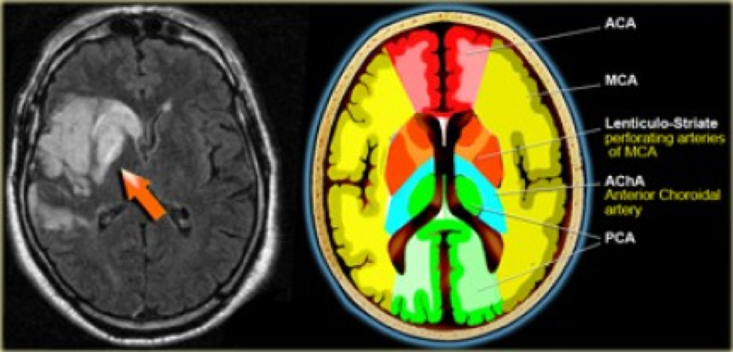
Middle Cerebral Artery W Radiology

Middle Cerebral Artery W Radiology The mca arises from the internal carotid artery as the larger of the two main terminal branches (the other being the anterior cerebral artery), coursing laterally into the lateral sulcus where it branches to perfuse the cerebral cortex. Advances in neuroimaging and endovascular therapy have further highlighted the clinical importance of this vessel, enabling targeted interventions for acute stroke management. this activity explores the mca’s anatomy, functional role, and clinical implications.

Middle Cerebral Artery W Radiology For these reasons, we present this anatomic review of the mca, reviewing its seg ments and anatomic limits, its branching patterns, and its anatomic variants. Objective: we aimed to investigate the geometric features of the middle cerebral artery (mca) and their relevance to plaque distribution and ischemic stroke. methods: we reviewed our institutional vessel wall imaging database. Aim: to assess whether the presence of the hyperintense middle cerebral artery (mca) sign, detected using magnetic resonance imaging (mri), has any prognostic value in subacute infarction. the results were also compared with computed tomography (ct). Future developments in high resolution mr imaging to depict intracranial atherosclerosis are explored in this review; these advances will guide endovascular therapy and the comparison of novel interventions.

Middle Cerebral Artery Anatomy Radiology Notes Aim: to assess whether the presence of the hyperintense middle cerebral artery (mca) sign, detected using magnetic resonance imaging (mri), has any prognostic value in subacute infarction. the results were also compared with computed tomography (ct). Future developments in high resolution mr imaging to depict intracranial atherosclerosis are explored in this review; these advances will guide endovascular therapy and the comparison of novel interventions. To review morphometry, morphology, branching patterns and anomalies of middle cerebral artery (mca). the databases of pubmed, google scholar and scopus were searched with different keywords. Horizontal part of the middle cerebral artery which gives rise to the lateral lenticulostriate arteries which supply most of the basal ganglia. the m2 segment is the part in the sylvian fissure and the m3 segment is the cortical segment. There are however certain features specific to middle cerebral artery infarct, and these are discussed below. for both ct and mri it is worth dividing the features according to the time course.
Comments are closed.