Solved 1 Describe The Difference Between Dark Field And Bright
Solved 1 Describe The Difference Between Dark Field And Bright Field Microscopy Course Optical microscopes employ visible light and a series of lenses to magnify the specimen and view it in detail. a bright field microscope uses light rays to create a dark image against a bright background. In this technique, light is directed at an angle, making it miss the objective lens and creating a dark background. the specimen appears bright against a dark background.
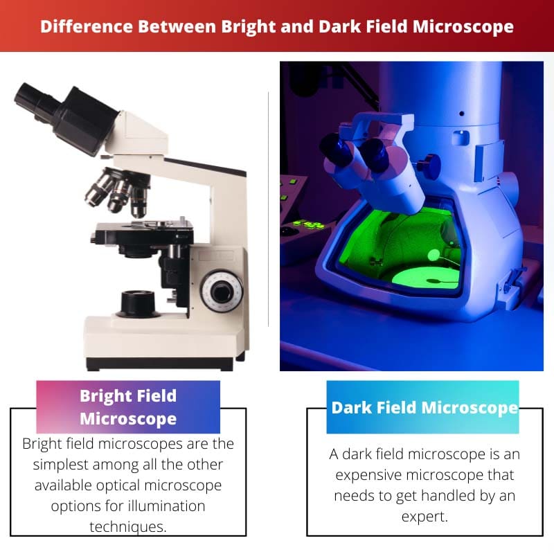
Solved 1 Describe The Difference Between Dark Field And Bright Field Microscopy Course In summary, bright field microscopy is suitable for observing stained or pigmented samples with lower contrast, while dark field microscopy is ideal for transparent, live, or unstained specimens, offering higher contrast and improved visibility of fine structures. Contrary to bright field, dark field illumination lights the specimen from the sides, ensuring that only the light scattered by the specimen enters the microscope’s objective. this results in a bright specimen on a dark background. They produce dark images on a bright background. dark field microscopes, on the other hand, are usually used in viewing live specimens due to their ability to increase contrast and resolution without the need for staining procedures. Bright field microscopy uses transmitted light to illuminate the sample, creating a bright background, while dark field microscopy scatters light around the specimen, resulting in a dark background.

Simulation Result Of Conversion Between Dark And Bright Field Download Scientific Diagram They produce dark images on a bright background. dark field microscopes, on the other hand, are usually used in viewing live specimens due to their ability to increase contrast and resolution without the need for staining procedures. Bright field microscopy uses transmitted light to illuminate the sample, creating a bright background, while dark field microscopy scatters light around the specimen, resulting in a dark background. Darkfield microscopy shows the specimens bright on a dark background. brightfield microscopes that have a condenser with a filter holder can be easily converted to darkfield by placing a patch stop filter into the filter holder. the filter blocks the direct light of the microscope. When you view a particular specimen under a bright field microscope, you will observe that the specimen is dark while its background is bright; hence the name bright field microscope. Describe the difference between dark field and bright field microscopy? 2. what are the major shapes of bacteria and their arrangements? 3. give 5 differences between eukaryote and prokaryotic cells? 4. what is the difference between the cell wall and the cell membrane? your solution’s ready to go!. Bright field image is the most common image generated with a tem. some areas of the sample can absorb or scatter electrons and appear darker, while other areas that transmit electrons appear brighter.

Bright Vs Dark Field Microscope Difference And Comparison Darkfield microscopy shows the specimens bright on a dark background. brightfield microscopes that have a condenser with a filter holder can be easily converted to darkfield by placing a patch stop filter into the filter holder. the filter blocks the direct light of the microscope. When you view a particular specimen under a bright field microscope, you will observe that the specimen is dark while its background is bright; hence the name bright field microscope. Describe the difference between dark field and bright field microscopy? 2. what are the major shapes of bacteria and their arrangements? 3. give 5 differences between eukaryote and prokaryotic cells? 4. what is the difference between the cell wall and the cell membrane? your solution’s ready to go!. Bright field image is the most common image generated with a tem. some areas of the sample can absorb or scatter electrons and appear darker, while other areas that transmit electrons appear brighter.

Bright Vs Dark Field Microscope Difference And Comparison Describe the difference between dark field and bright field microscopy? 2. what are the major shapes of bacteria and their arrangements? 3. give 5 differences between eukaryote and prokaryotic cells? 4. what is the difference between the cell wall and the cell membrane? your solution’s ready to go!. Bright field image is the most common image generated with a tem. some areas of the sample can absorb or scatter electrons and appear darker, while other areas that transmit electrons appear brighter.
Comments are closed.